Endocrine Pathology Slide Seminar
Vania Nosé, MD, PhD – Professor of Pathology, Harvard Medical School, Massachusetts General Hospital, Boston, MA, USA
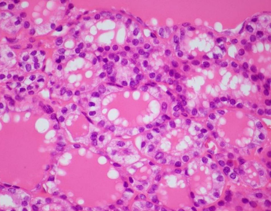
Case 1
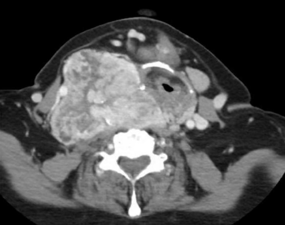
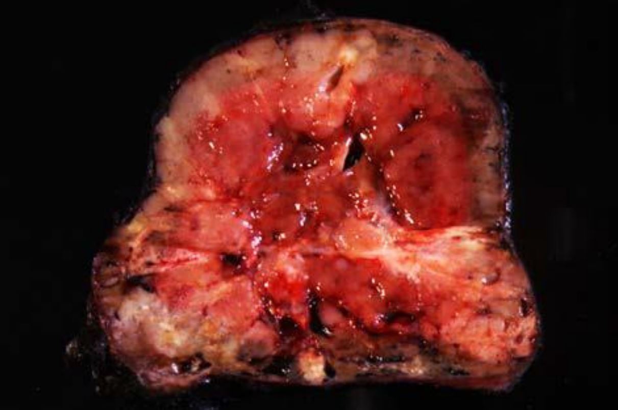
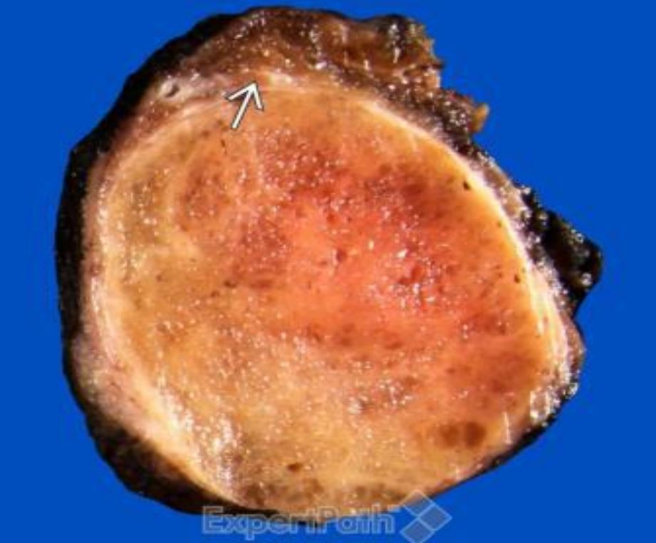
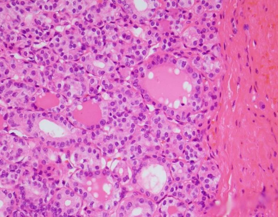
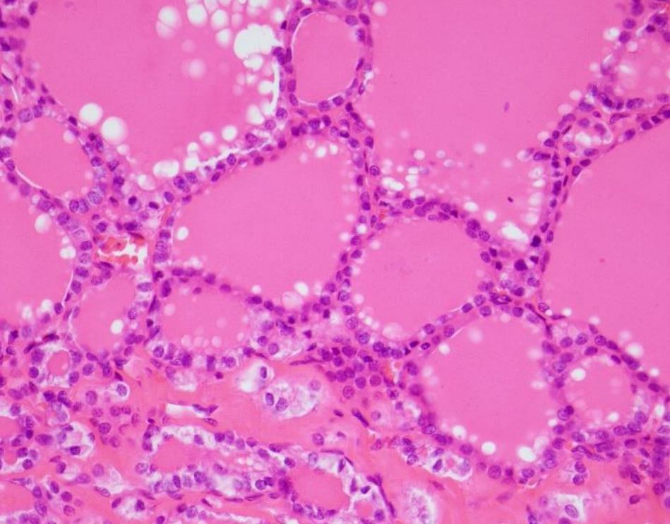
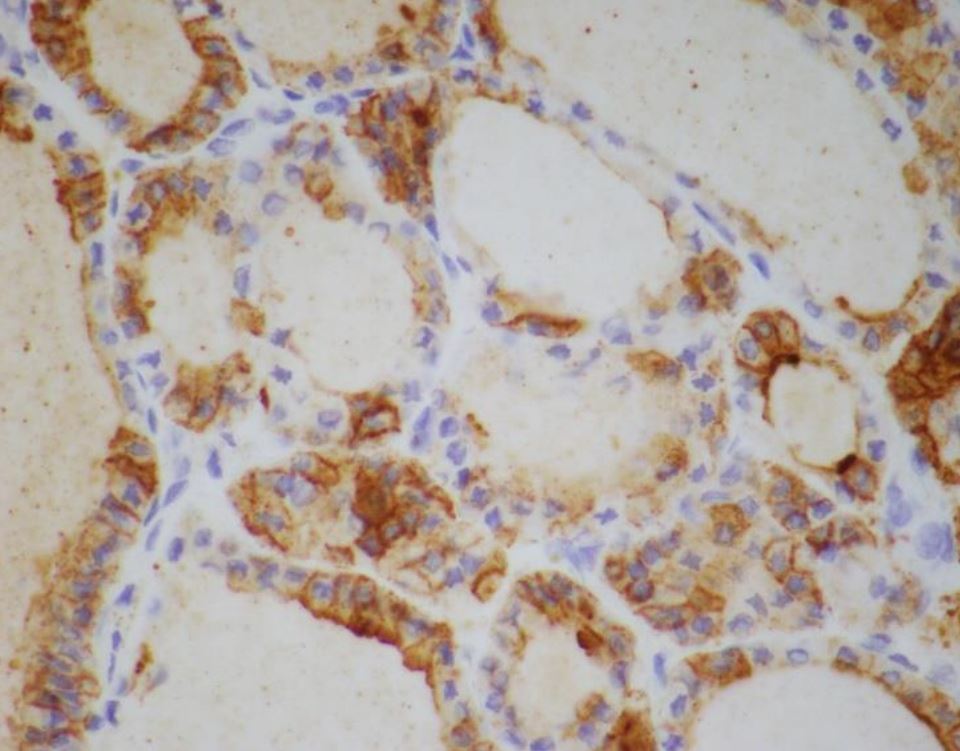
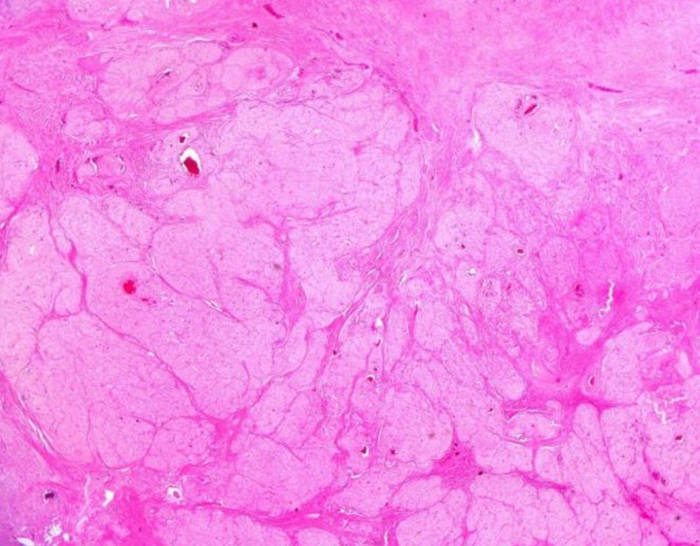
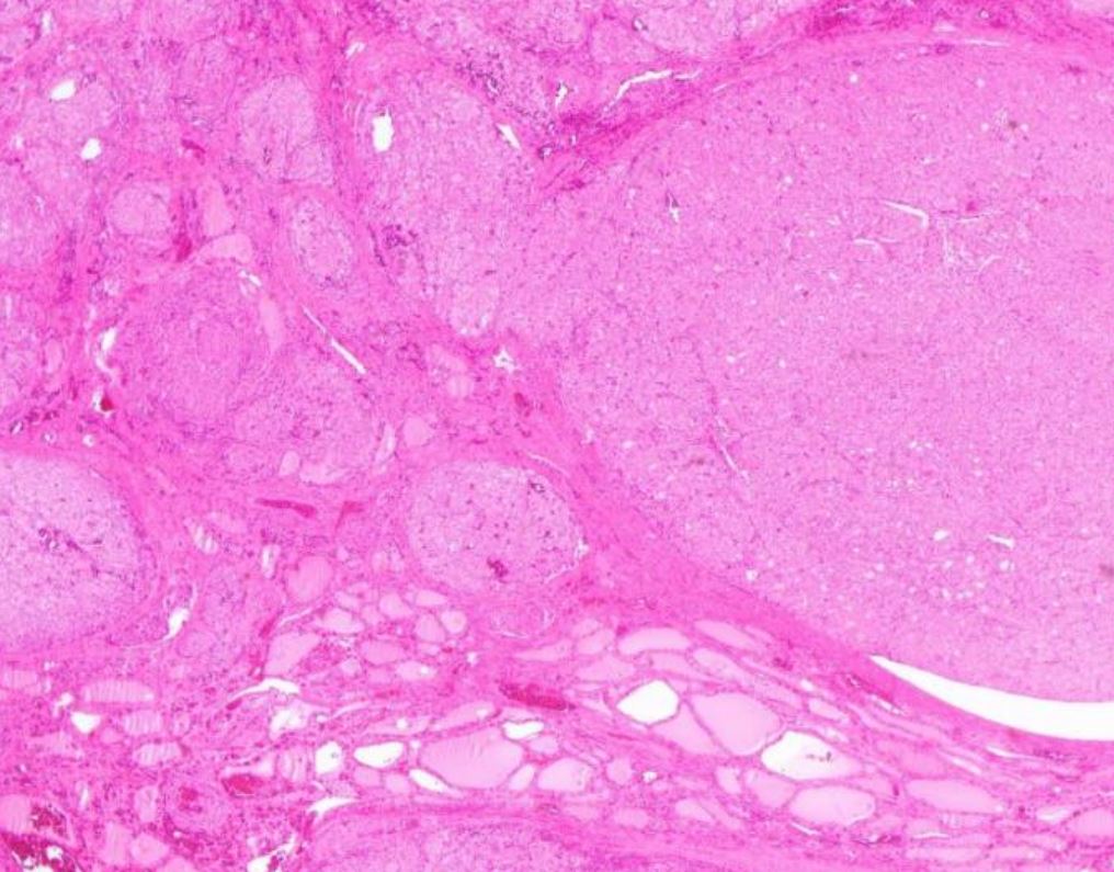
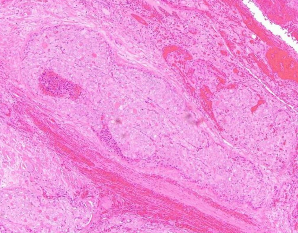
75-year-old woman with slowly enlarging thyroid followed for several years, with a large goiter on the right side she also has HPT with a calcium of 10.9 and PTH of 93 with a possible parathyroid adenoma adjacent to the left that is the smaller side thyroid. CT scan and ultrasound shows a right lobe which is mass of 9 cm going from the parotid to just below the clavicle. She has several small nodules in the left lobe as well. FNA were done in several occasions, benign.
-
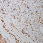
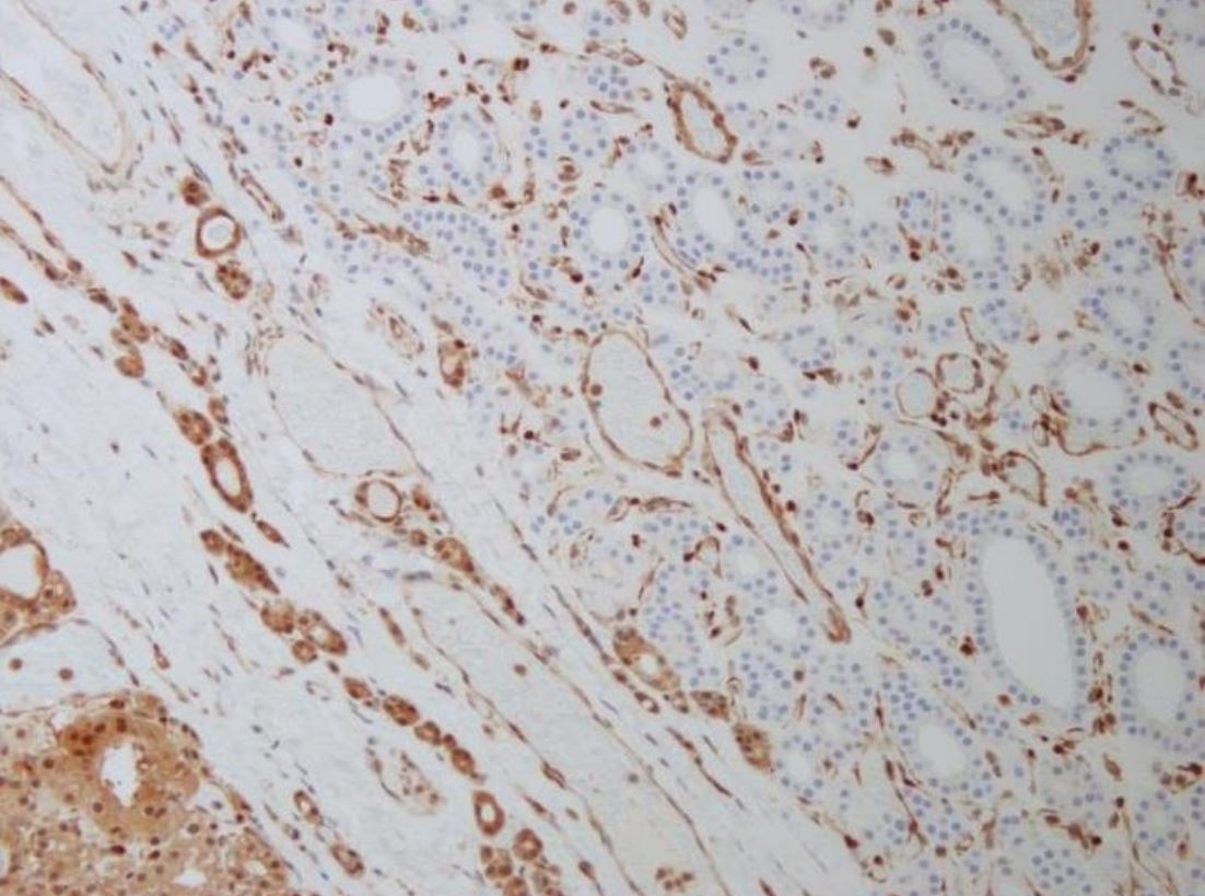
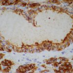
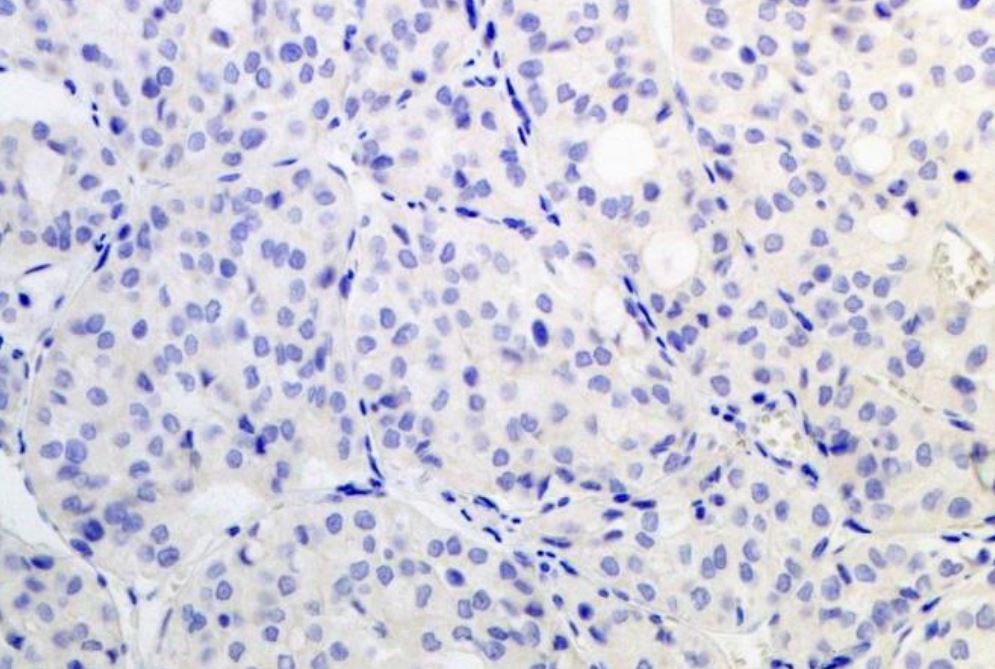
- PTEN IHC
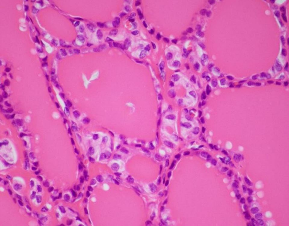
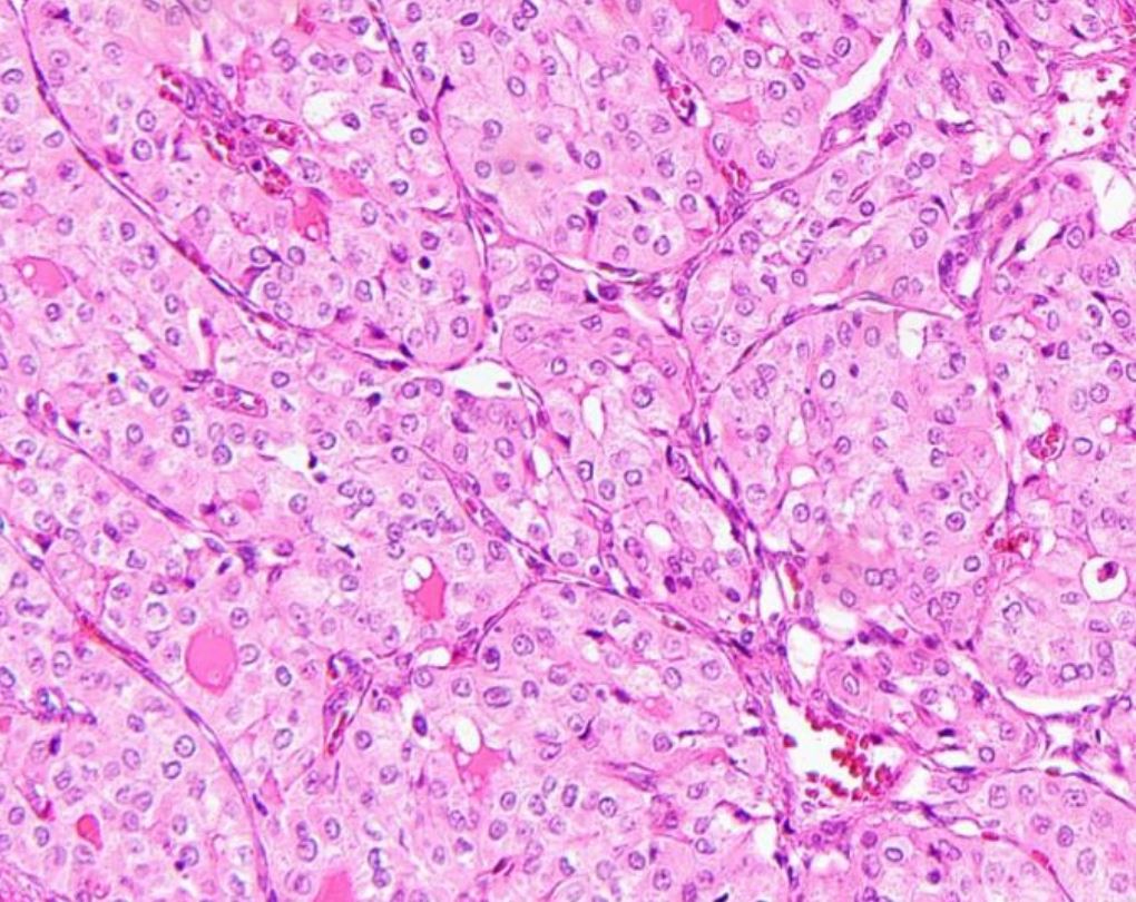
Case 2
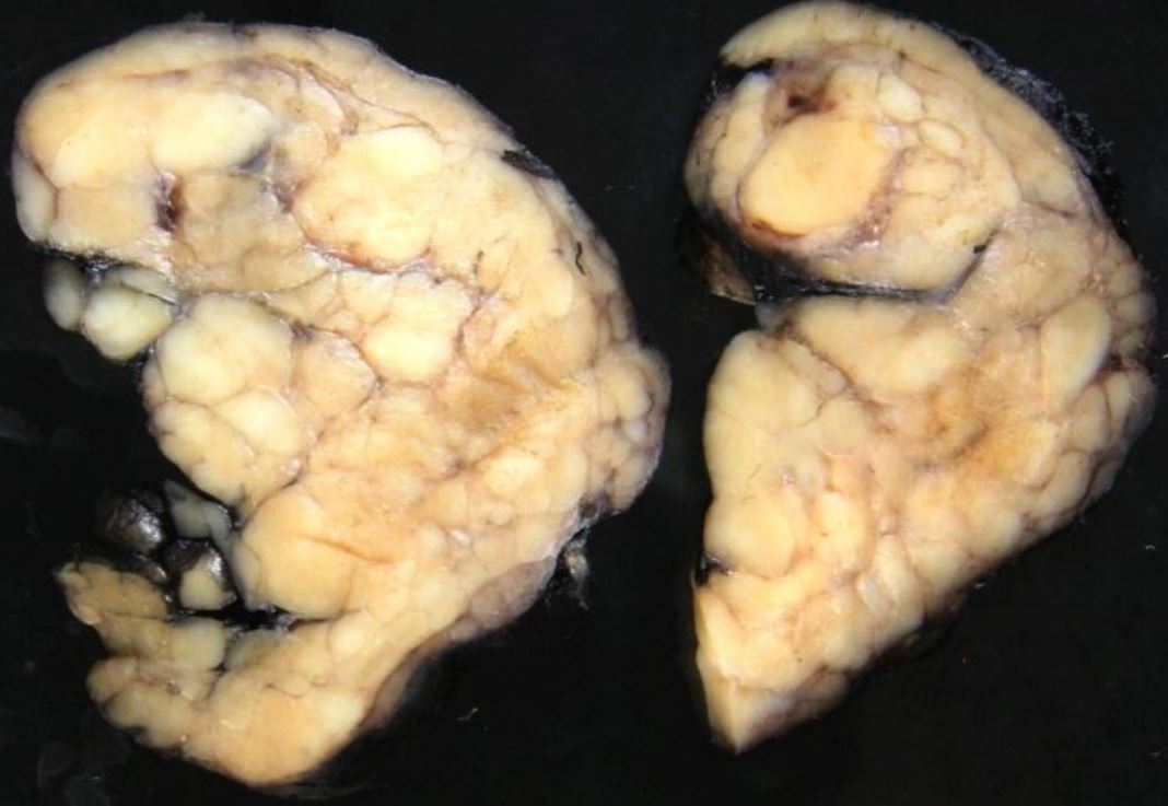
54 y.o. female comes for consultation about treatment of her large, symptomatic, right thyroid nodule with FNA cytology showing a follicular neoplasm with positive NRAS mutation. Thyroseq suggest an 80% risk of associated thyroid malignancy.
-
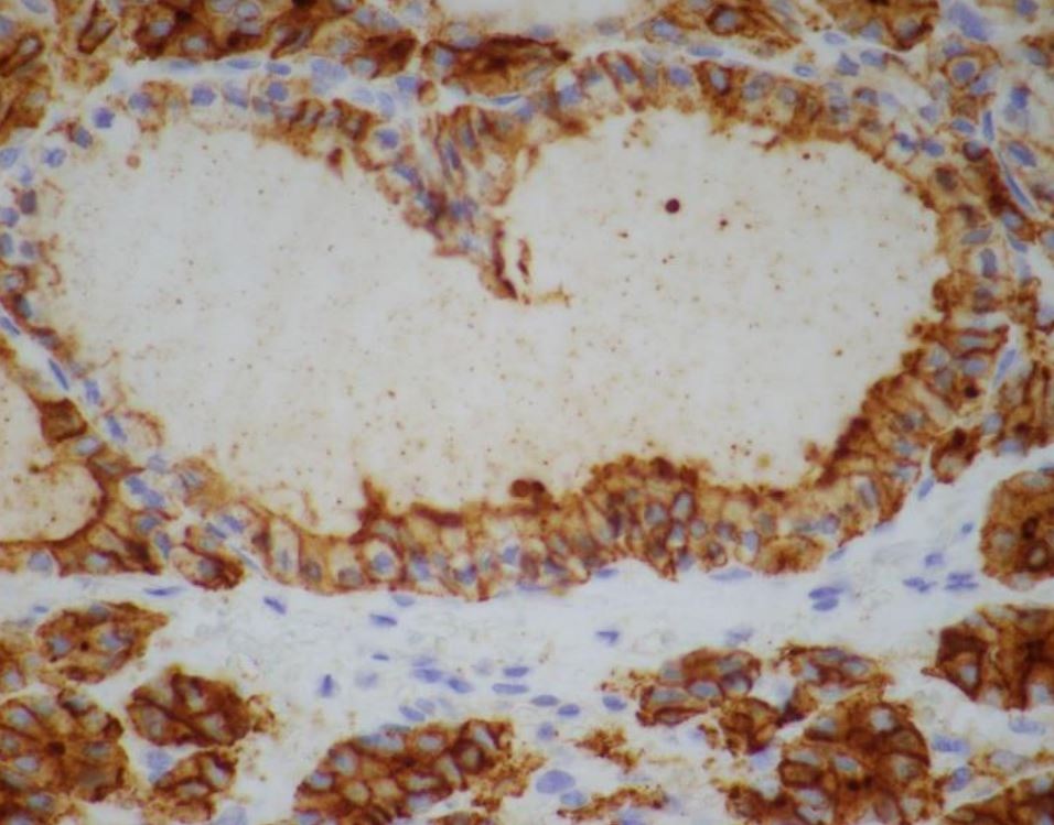
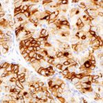
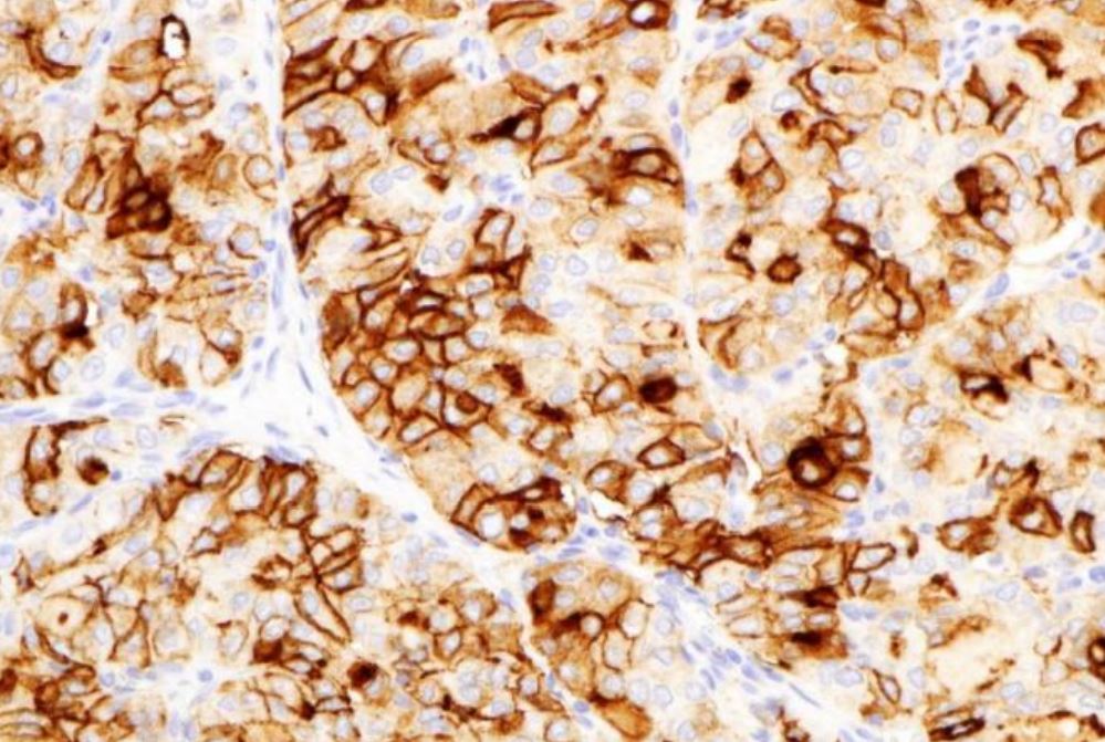
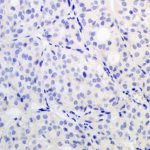
- HBME1 IHC
Case 3
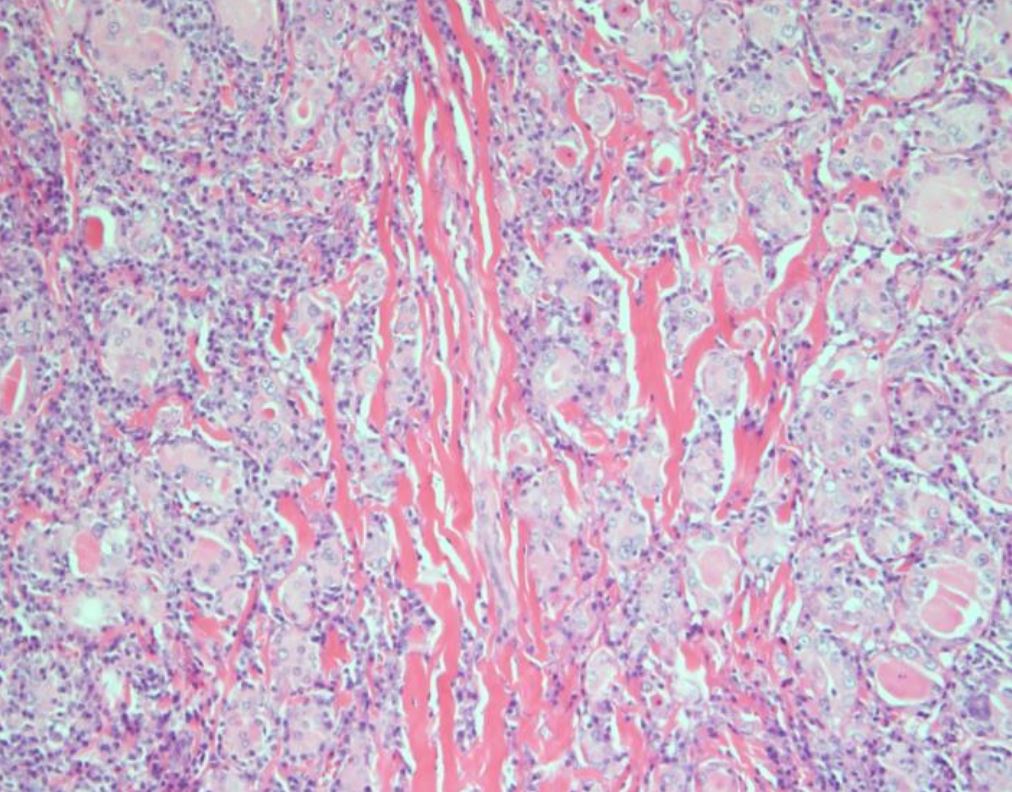
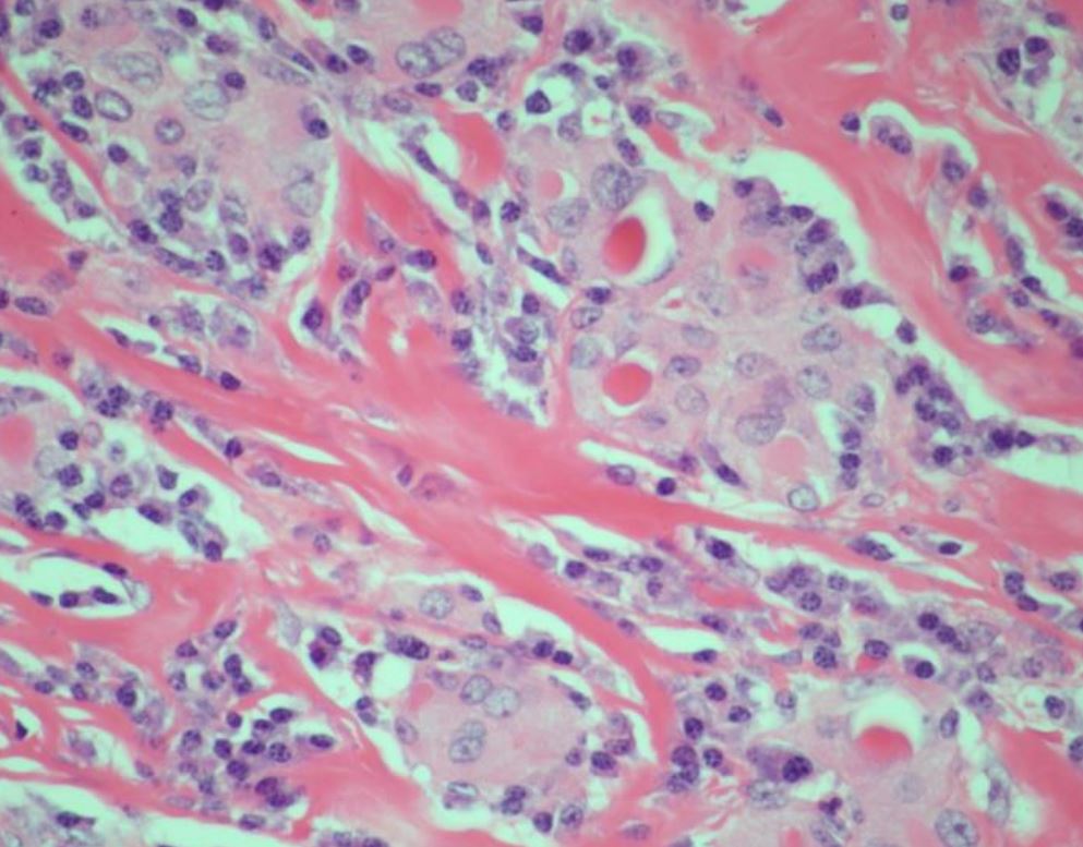
42-year-old female music teacher presented to her primary care provider after experiencing progressive throat tightness over the preceding year. She also noticed an anterior neck mass and bilateral cervical lymph nodes, which have been gradually enlarging.
Cervical sonography and computer tomography both revealed a 5.4 x 5.1 x 4.3 cm mass replacing the entire right thyroid lobe with extension into the isthmus. Also noted was bilateral central neck adenopathy and right lateral neck adenopathy. Fine needle aspiration was positive for papillary thyroid carcinoma.
-
- HBME1 IHC
-
- BRAF IHC
Case 4
62 ear old woman with long history of Hashimoto thyroiditis, at least for 20 years, when a biopsy was performed that was consistent with Hashimoto. She has been followed since, and the repeated ultrasound has been heterogeneous, the right lobe is larger than left, but with no nodules. TPO antibody of 905 and TSH of 3.259.
-
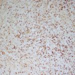
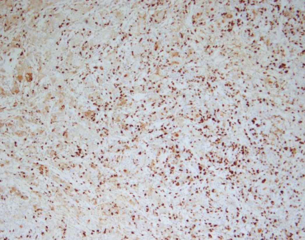
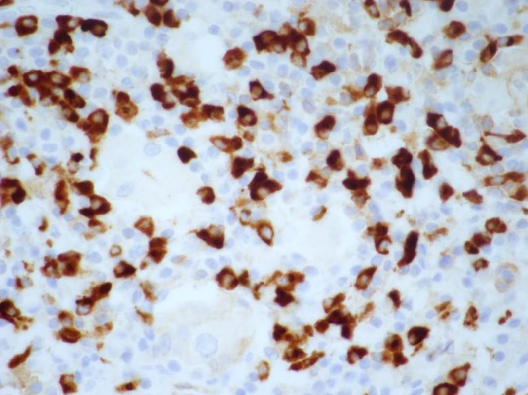
- IgG4


 Bourbon Resort Cataratas do Iguaçu
 Av. das Cataratas, 2345 - Vila Yolanda,